











Ultrasound of the Scrotum (Testicles) at Reprolife: Your Path to Accurate Diagnosis of Men’s Health
Ultrasound (US) examination of the scrotum (testicles) is a fundamental method in andrology and urology, providing non-invasive, painless, and highly informative diagnostics of the condition of the testicles, epididymis, and spermatic cords. At Reprolife center, we use ultrasound not only to confirm obvious pathologies but also as a key tool in the diagnosis of male infertility and the prevention of serious diseases.
Advantages of Scrotal Ultrasound at Our Center
-
Safety and Non-Invasiveness: The procedure does not use ionizing radiation and can be performed repeatedly without harm to health.
-
High Resolution: Modern equipment allows the visualization of structures down to a millimeter in size.
-
Blood Flow Assessment (Doppler): The mandatory use of color Doppler ultrasonography to assess the speed and direction of blood flow, which is critical for diagnosing varicocele and testicular torsion.
-
Expert Approach: The examinations are conducted by diagnostic physicians specializing in reproductive and urological pathology.
When is Scrotal Ultrasound Necessary? Key Indications
Scrotal ultrasound is a mandatory examination, prescribed by a urologist or andrologist in the following cases:
-
Symptoms and Acute Conditions:
-
Scrotal Pain: Acute, sudden, or chronic, dull pain.
-
Swelling and Redness: Signs of inflammation (orchitis, epididymitis).
-
Palpable Masses: Sensation of lumps, nodules, cysts, or enlargement of one of the testicles.
-
Trauma: Examination after an impact or injury to rule out a hematoma or testicular rupture.
-
-
Diagnosis of Infertility and Structural Anomalies:
-
Male Infertility: It is the standard in initial diagnostics for identifying hidden causes.
-
Varicocele: Diagnosis of the enlargement of the veins of the spermatic cord, which is often the cause of impaired spermatogenesis.
-
Cryptorchidism: Ultrasound helps to localize testicles that have not descended into the scrotum.
-
Post-Operative Assessment: Monitoring the condition after surgical treatment for varicocele or other interventions.
-
-
Detection of Fluid Collections:
-
Hydrocele: Accumulation of fluid (water on the testicle).
-
Spermatocele/Epididymal Cysts: Diagnosis of benign fluid-filled formations.
-
What Can Scrotal Ultrasound with Doppler Diagnose?
The use of B-mode ultrasound (for structure) and Doppler (for blood flow) provides a complete diagnostic picture:
-
Testicular Torsion (Torsio): Doppler ultrasound is the only rapid method for assessing blood flow, which helps differentiate torsion (lack of blood flow) from inflammation.
-
Testicular Tumors: Accurate determination of the size, location, and structure of suspicious neoplasms, which is critical for the early initiation of oncological treatment.
-
Varicocele: Determination of the degree of vein enlargement and the presence of reflux (reverse blood flow) during the Valsalva maneuver.
-
Inflammatory Processes: Assessment of the size and structure of the testicle/epididymis in orchitis and epididymitis.
-
Seminal Duct Obstruction: Signs of blockage that may be the cause of azoospermia.
How is the Ultrasound Procedure Performed?
-
Preparation: No special preparation is required. Standard hygiene procedures are sufficient.
-
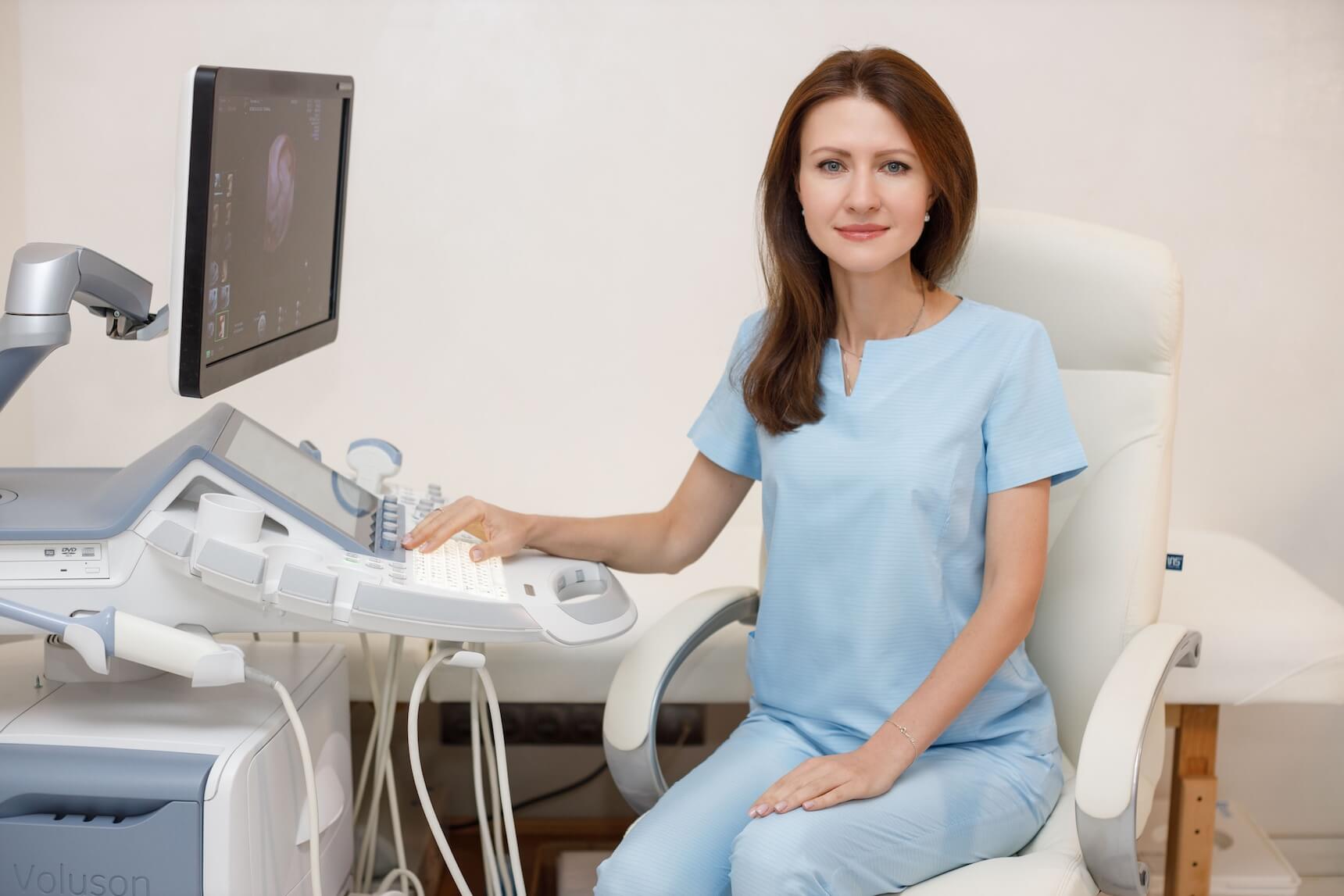
Procedure: The patient lies on their back. A hypoallergenic gel is applied to the scrotal skin. The doctor gently moves the ultrasound transducer, visualizing the structures in real time.
-
Doppler Ultrasonography: Doppler can be performed simultaneously to assess the speed and direction of blood flow, which is critically important for diagnosing varicocele and testicular torsion.
-
Duration: The examination is fast and usually takes 15–30 minutes.
-
Result: The patient receives a detailed conclusion and images immediately after the procedure is completed.
-
Important: In case of suspicion of acute conditions (testicular torsion, acute epididymitis), the ultrasound is performed immediately.
Why Choose Reprolife for Scrotal Ultrasound?
In our center, you receive not just an image, but expert diagnostics integrated into the overall treatment plan:
-
Focus on Reproductive Medicine: Our specialists have a deep understanding of how detected pathologies (e.g., varicocele) affect sperm quality and fertility.
-
Comprehensive Approach: Ultrasound results are immediately analyzed in the context of the patient’s spermogram and hormonal status.
-
Discretion: We guarantee complete confidentiality, comfort, and attentive care for the patient.
Early diagnosis is the key to successful treatment! Do not delay taking care of your health. Accurate diagnosis is the first step towards successful treatment and realizing parenthood plans. Schedule your scrotal ultrasound today!